
Cranial cruciate ligament tear

Cranial cruciate ligament (CCL) rupture is one of the most common orthopedic conditions seen in veterinary practice and is the most common cause of acute (sudden) hindlimb lameness in dogs. The CCL is one of two major ligaments that provides stability between the femur and tibia in the canine stifle (knee), similar to the anterior cruciate ligament (ACL) in humans. The CCL functions to prevent cranial (forward) translation of the tibia. The CCL can be thought of as a rope composed of many individual fibers. Over time, these fibers can gradually weaken and break down through a degenerative process. Once enough fibers have been damaged, the stifle becomes unstable, ultimately resulting in a CCL rupture. Genetic, environmental, and conformational factors can all contribute to the development of CCL disease. CCL ruptures can occur in dogs of any breed, size, or activity level.
Typical clinical signs of a CCL rupture include weight-bearing lameness, off-loading the affected limb while standing, and external rotation and extension of the affected limb when sitting. During an orthopedic examination, findings may include stifle effusion (increased joint fluid), pain on extension of the stifle, a medial buttress (thickening of the soft tissues on the medial aspect of the joint) in chronic cases, excessive internal rotation, and positive cranial drawer and tibial thrust tests.
These tests may demonstrate abnormal movement or subluxation of the tibia relative to the femur. The medial meniscus is also commonly injured concurrently with a CCL rupture. This is largely due to the anatomical attachment of the medial meniscus to the tibia, which can result in the meniscus becoming compressed or crushed when there is abnormal cranial advancement of the tibia.
Although several surgical procedures can be performed to stabilize a CCL-deficient stifle, the most commonly recommended procedure is a Tibial Plateau Leveling Osteotomy (TPLO). This surgery first starts with evaluation of the knee joint to examine the cruciate ligaments as well as both menisci. We recommend that this is done arthroscopically, though it can be done through a small “open” approach. If the meniscus is injured, the damaged area is removed. Next, the TPLO is performed. During a TPLO, a semicircular cut is made through the proximal (upper) tibia, and the tibial plateau is rotated into a relatively level orientation. This alters the biomechanics of the stifle and creates a stable configuration that does not rely on the CCL to prevent cranial translation of the tibia.
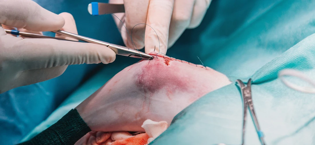
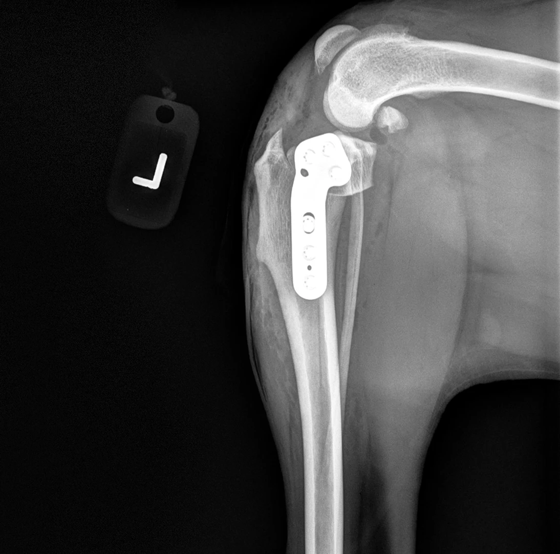
The osteotomy is then stabilized with a bone plate and screws. The recovery period following TPLO surgery generally lasts approximately 8–12 weeks. During this time, patients should be strictly confined and permitted only controlled rehabilitation exercises designed to maintain and strengthen muscle mass while gradually returning the limb to full function.
CCL rupture is also a primary contributor to the development of degenerative joint disease (osteoarthritis) in the stifle. TPLO surgery can slow the progression of osteoarthritis and help patients remain comfortable and active for a longer period of time. One of the most significant long-term concerns, however, is that once a CCL rupture has occurred in one stifle, there is an estimated 40–60% chance that the other stifle will also develop CCL disease at some point, with many cases occurring within two years of the initial rupture. Fortunately, once both stifles have been treated surgically, there are no remaining knees at risk for developing a CCL rupture!