
Medial Patella Luxation

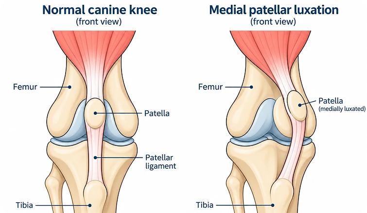
Patella luxation is one of the most common orthopedic conditions in dogs. Medial patellar luxation primarily affects small breeds, including Boston and Yorkshire terriers, Chihuahuas, Pomeranians, and miniature poodles. While large-breed dogs can also develop medial patellar luxation, they more often develop lateral patellar luxation. The patella, or kneecap, is a small bone embedded within the tendon of the quadriceps muscles of the thigh. Normally, the patella glides within a groove in the femur (thigh bone) as the knee flexes. The patellar tendon attaches to the tibial crest, a bony prominence on the tibia (shin bone) just below the knee. Together, the quadriceps muscle, patella, and patellar tendon form the extensor mechanism, which is normally well aligned.
Patellar luxation (dislocation) occurs when the patella moves outside the femoral groove during knee flexion. It is classified as medial or lateral, depending on whether the patella moves toward the inside or outside of the knee, respectively. In most dogs, patellar luxation is developmental and has a genetic component. The femoral groove is often shallow or absent, and the tibial crest may be positioned abnormally, resulting in misalignment of the entire extensor mechanism. In severe cases, malformation of the femur and/or tibia may also contribute to the luxation.
Clinical signs vary considerably depending on the grade and severity of the luxation. Common signs include a “skipping” gait, intermittently carrying the affected limb, or shaking or extending the limb before returning it to normal use. In more severe cases, dogs may develop a pronounced bow-legged appearance of the hind limbs. Patellar luxation may also be an incidental finding during a routine physical examination. Repeated luxation can damage and erode the cartilage within the knee, eventually exposing underlying bone and resulting in osteoarthritis and pain.
Physical examination findings depend on the severity of the luxation:
At Kin, we offer a range of minimally invasive procedures. The best approach depends on your pet’s condition, anatomy, and overall health.
- Grade I: The patella can be manually luxated but returns to its normal position when released. Dogs are generally not lame, and the condition is often identified incidentally.
- Grade II: The patella luxates intermittently, commonly causing an occasional “skipping” gait while walking or running.
- Grade III: The patella is luxated most or all of the time but can be manually reduced. Lameness ranges from occasional skipping to weight-bearing lameness.
- Grade IV: The patella is permanently luxated and cannot be manually reduced. Dogs typically walk with their hindquarters crouched because they cannot fully extend the knee.
The decision between conservative and surgical treatment depends on the grade of luxation, clinical signs, progression, and presence of osteoarthritis. Depending on the individual anatomy and severity of the condition, one or more surgical procedures may be necessary:
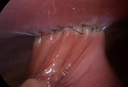
- Soft-tissue reconstruction: The tissues surrounding the patella are adjusted to release tension on the side toward which the patella luxates and tighten the opposite side. This may include releasing contracted tissues or performing an imbrication of loose tissues.
- Trochlear deepening: The femoral groove is deepened to allow the patella to seat securely in its normal position. Several techniques can be used to achieve this.
- Tibial tuberosity transposition: The tibial tuberosity, where the patellar tendon attaches, is repositioned to realign the extensor mechanism and secured with pins or a pin-and-tension-band construct.
- Femoral corrective osteotomy: In severe cases involving abnormal femoral angulation or torsion, the femur may be surgically cut and realigned, then stabilized with a bone plate.