
Intervertebral Disc Disease (IVDD)
Olby NJ, Moore SA, Brisson B, Fenn J, Flegel T, Kortz G, Lewis M, Tipold A. ACVIM consensus statement on diagnosis and management of acute canine thoracolumbar intervertebral disc extrusion. J Vet Intern Med.2022;36(5):1570–1596. doi:10.1111/jvim.16480.
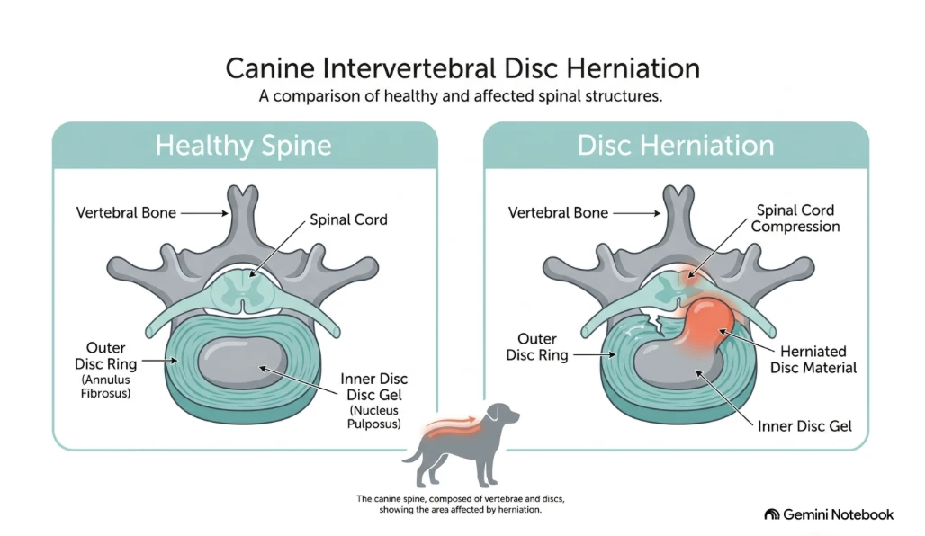
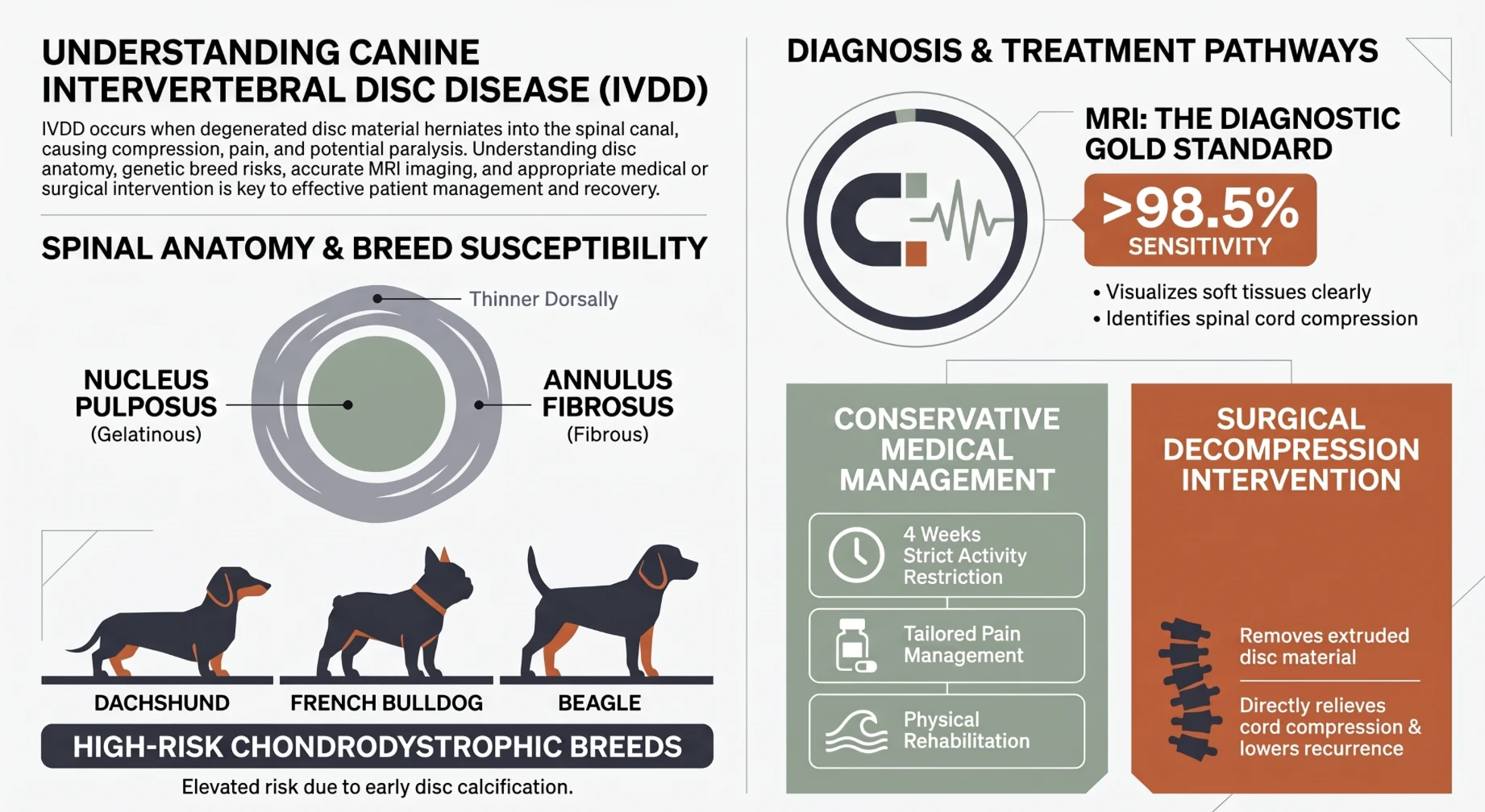
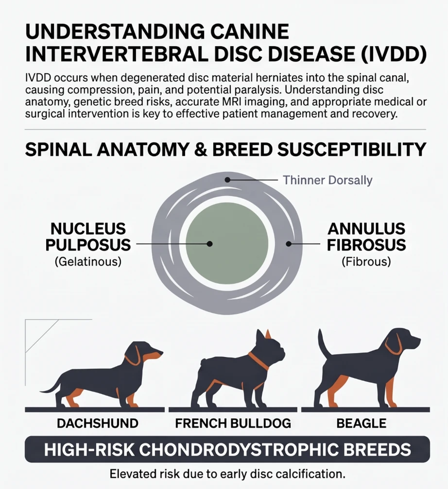
Intervertebral discs are shock-absorbing cushions between the bones of the spine (vertebrae). These consist of an outer fibrous ring (called the annulus fibrosus) and a central gelatinous center (called the nucleus pulposus). The inner nucleus pulposus is rich in water (approximately 80%) and proteoglycans to absorb mechanical stress, while the surrounding annulus fibrosus is made of concentric fibrous layers.
Intervertebral Disc Disease (IVDD) is the general term for degeneration of the intervertebral disc. This manifests in two primary forms depending on the dog’s breed and underlying pattern of disc degeneration. Breeds—such as Dachshunds, French Bulldogs, Beagles, and Spaniels—are strongly predisposed to an acute disc herniation (Hansen Type I extrusion) due to early disc dehydration, cartilage transformation, and calcification. This change results in the gelatinous core of the disc becoming a thick paste or a firm pebble that is under constant pressure. When this material breaks through the outer fibrous layer (the annulus fibrosus, is thinner on its upper side) and ruptures into the spinal canal, this is what is called an Intervertebral Disc Herniation (IVDH). French Bulldogs in particular often present with disc herniations at younger ages and can be at a higher risk for severe spinal cord complications. In contrast, larger dogs (such as German Shepherds or Labradors) more commonly develop Hansen Type II protrusion, a slower, age-related degeneration where the disc gradually bulges into the spinal canal over time.
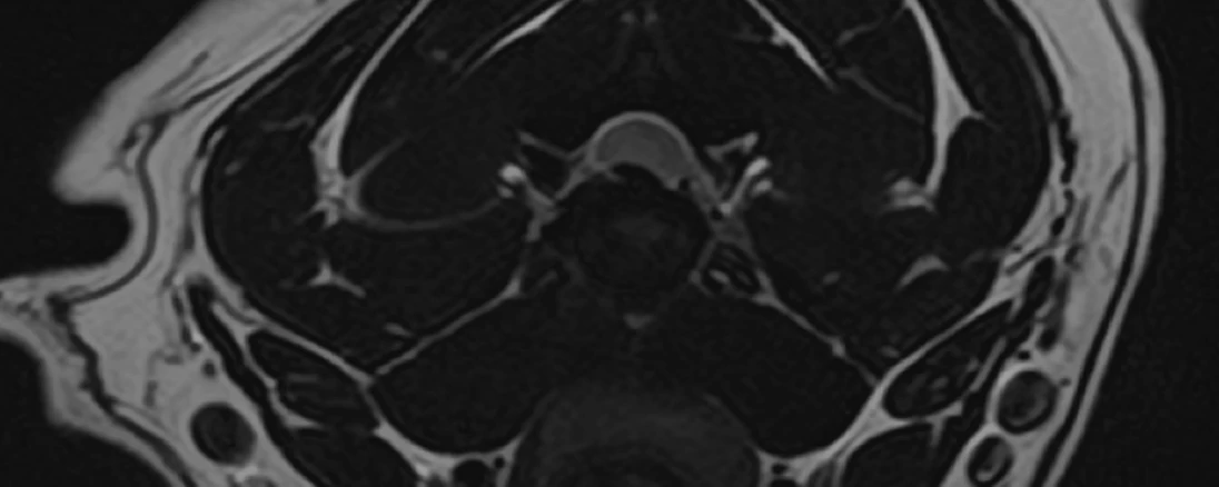
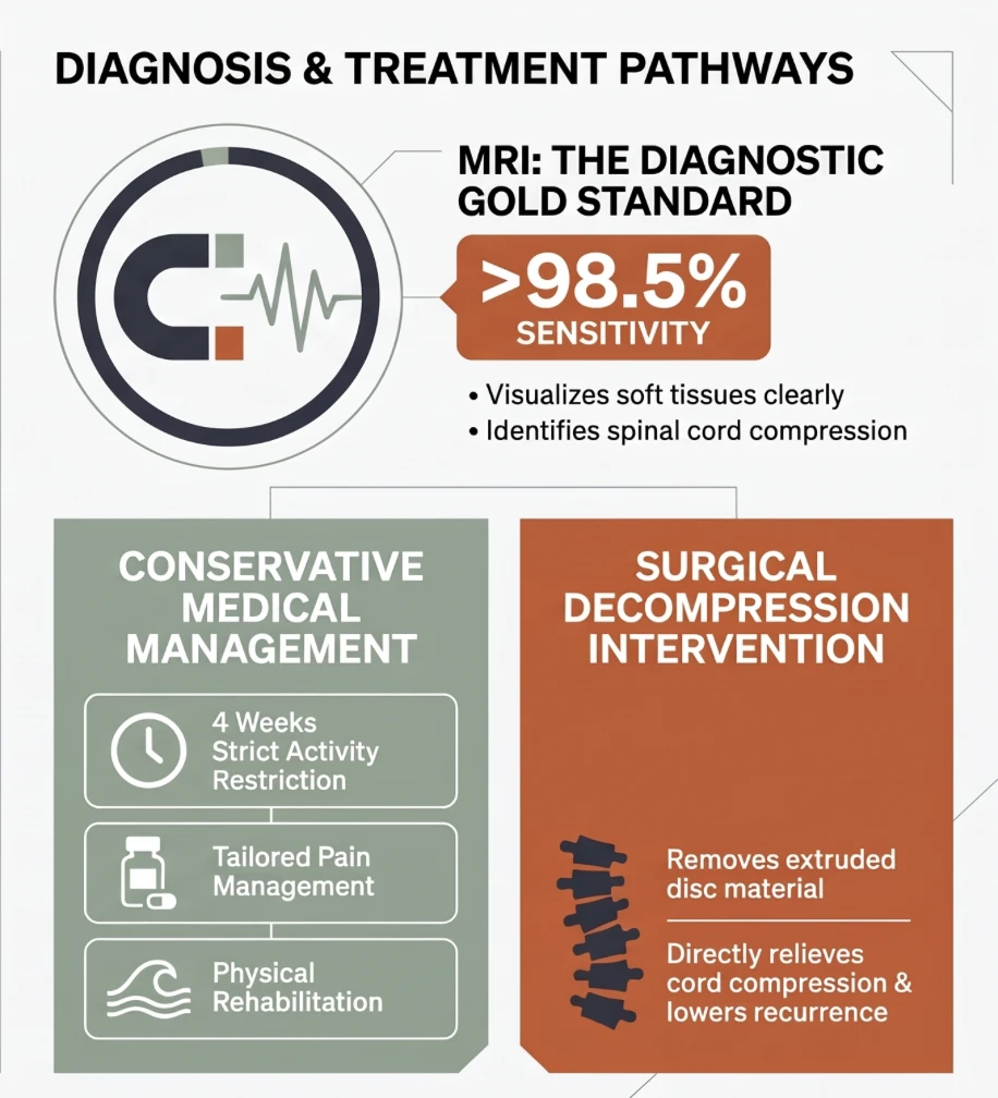
Magnetic Resonance Imaging (MRI) is the premier diagnostic tool for evaluating intervertebral disc disease, offering a diagnostic accuracy exceeding 98.5%. MRI provides detailed soft-tissue visualization that clearly displays the extruded or protruding disc material compressing the spinal cord. Furthermore, MRI enables veterinarians to assess for secondary spinal cord damage—such as inflammation, swelling, or fluid accumulation and other diseases which can cause similar clinical signs.
Treatment for IVDD is categorized into medical or surgical management based on the severity of the dog’s neurological deficits. Medical (conservative) management is appropriate for dogs with mild pain or preserved walking ability; it requires approximately 4 weeks of strict cage rest to allow the outer disc wall to heal, paired with anti-inflammatory medications (such as NSAIDs) and basic rehabilitation exercises. Surgical management (such as a hemilaminectomy in the lower back or a ventral slot in the neck) is indicated for dogs that are unable to walk, experiencing severe pain, or failing conservative (medical) treatment. Surgery physically removes the herniated material to relieve spinal cord compression and often includes disc fenestration to minimize the risk of future disc recurrences.
Following IVDD surgery, your dog will require 4 to 6 weeks of strict crate or pen confinement to protect the surgical site, prevent spinal stress, and allow delicate tissues to heal properly. Outdoor activity must be limited strictly to short, leash-guided bathroom breaks, utilizing a supportive belly sling and chest harness to assist weak legs and prevent dangerous slips on slick flooring. The Kin Neurology team equips owners with the tools to accommodate post-operative rest including a sling and medications to help facilitate pain relief and mild anxiolysis.
We often joke that the bladder is the fifth and most important leg! Therefore, with loss of movement in the legs, it is critical to monitor bladder function—ensuring your dog is voluntarily voiding urine rather than leaking. Prior to discharge, if warranted based on your dog’s neurologic status, the Kin Neurology team will trial bladder expression with you so you feel comfortable prior to taking your pet home. We know managing a pet unable to walk on their own is stressful, so we are here to answer any questions or concerns you have along the way!